GHRH Receptor Biology and Gs-cAMP-PKA Signal Transduction
Growth hormone-releasing hormone (GHRH) is a 44-amino acid hypothalamic peptide that serves as the primary physiological stimulus for growth hormone (GH) synthesis and secretion from anterior pituitary somatotroph cells. The biological effects of GHRH are mediated through the GHRH receptor (GHRH-R), a 423-amino acid class B (secretin family) G-protein coupled receptor encoded by the GHRHR gene on human chromosome 7p14. The receptor was first cloned from pituitary tissue by Mayo (1992), and its molecular pharmacology has since been extensively characterized in transfected cell systems and primary pituitary cultures (Mayo KE. Mol Endocrinol. 1992;6(10):1734-1744. doi:10.1210/mend.6.10.1333056).
The GHRH-R extracellular N-terminal domain (ECD) adopts the characteristic alpha-beta-beta-alpha fold conserved across class B GPCRs, forming a hydrophobic binding groove that accommodates the amphipathic alpha-helix of GHRH. Residues 1-29 of GHRH contain the full determinants for receptor binding and activation, as demonstrated by the equipotent activity of GHRH(1-29)-NH2 (sermorelin) and full-length GHRH(1-44)-NH2 in cAMP accumulation assays (Ling N, et al. Biochem Biophys Res Commun.1984;122(1):304-310. doi:10.1016/0006-291X(84)90474-2). The N-terminal tyrosine (Tyr1) is critical for receptor activation: truncation or modification at this position abolishes agonist activity while retaining receptor binding affinity, producing competitive antagonist analogs.
Upon GHRH binding, the GHRH-R undergoes a conformational transition that promotes coupling to the stimulatory G-protein alpha subunit (Gs-alpha). Gs-alpha activates adenylate cyclase, catalyzing the conversion of ATP to cyclic AMP (cAMP). The resulting elevation in intracellular cAMP concentration activates two primary effector pathways. Protein kinase A (PKA), a serine/threonine kinase, is activated by cAMP binding to its regulatory subunits, releasing the catalytic subunits that phosphorylate downstream targets including CREB (cAMP response element-binding protein). Phosphorylated CREB translocates to the nucleus and binds CRE elements in the GH gene promoter, driving GH gene transcription.
The second cAMP effector, exchange protein directly activated by cAMP (Epac), has been identified as an additional mediator of GHRH-R signaling in somatotroph models. Epac activates the small GTPase Rap1, which contributes to calcium-independent exocytosis of GH-containing secretory granules (Bhatt DK, et al. J Mol Endocrinol. 2012;49(3):R1-R10. doi:10.1530/JME-12-0095). The relative contributions of PKA- and Epac-dependent pathways to GHRH-stimulated GH release have been dissected using pathway-selective cAMP analogs (8-Br-cAMP for PKA, 8-pCPT-2'-O-Me-cAMP for Epac) in primary rat pituitary cell cultures.
GHRH-R signaling also involves cross-talk with ion channel activity. PKA-mediated phosphorylation of voltage-gated potassium channels (Kv) and L-type calcium channels (Cav1) modulates membrane excitability in somatotroph cells. Inhibition of Kv channels depolarizes the cell membrane, facilitating calcium entry through voltage-gated calcium channels and triggering GH vesicle exocytosis. This electrophysiological component of GHRH-R signaling has been characterized using patch-clamp recordings in primary pituitary somatotroph preparations and GH-secreting cell lines such as GH3 and MtT/S cells.
GHS-R1a Pharmacology and the Gq-PLC-IP3 Signaling Pathway
The growth hormone secretagogue receptor type 1a (GHS-R1a) is a 366-amino acid class A (rhodopsin-like) GPCR that serves as the cognate receptor for ghrelin, a 28-amino acid octanoylated peptide hormone primarily secreted by X/A-like enteroendocrine cells of the gastric oxyntic mucosa. The receptor was identified as a target for synthetic growth hormone-releasing peptides (GHRPs) before ghrelin itself was discovered, and was originally cloned by Howard et al. in 1996 (Howard AD, et al. Science.1996;273(5277):974-977. doi:10.1126/science.273.5277.974).
Unlike GHRH-R, which primarily engages Gs proteins, GHS-R1a couples predominantly to Gq/11 proteins. Activation of Gq/11 stimulates phospholipase C-beta (PLC-beta), which hydrolyzes phosphatidylinositol 4,5-bisphosphate (PIP2) to generate two second messengers: inositol 1,4,5-trisphosphate (IP3) and diacylglycerol (DAG). IP3 binds to IP3 receptors on the endoplasmic reticulum, triggering the release of calcium from intracellular stores. DAG, together with the elevated intracellular calcium, activates protein kinase C (PKC), which phosphorylates a distinct set of substrates compared to those targeted by PKA downstream of GHRH-R.
The calcium mobilization triggered by GHS-R1a activation has been extensively characterized using fluorometric imaging plate reader (FLIPR) assays in GHS-R1a-expressing cell lines. In these assays, ghrelin and synthetic GHS-R1a agonists produce rapid, dose-dependent increases in intracellular calcium concentration ([Ca2+]i), with EC50 values in the low nanomolar range for potent agonists such as ipamorelin, GHRP-6, and GHRP-2. The calcium signal can be dissected into IP3-dependent release from intracellular stores (sensitive to thapsigargin pre-treatment) and subsequent store-operated calcium entry (SOCE) through plasma membrane channels.
A distinctive pharmacological property of GHS-R1a is its high level of constitutive activity. The receptor exhibits approximately 50% of maximal G-protein activation in the absence of any agonist ligand, as demonstrated by elevated basal IP3 turnover and constitutive internalization in heterologous expression systems. This constitutive activity is mediated by a conserved aromatic cluster (Phe279, Trp276, Phe312) in the transmembrane domain and has been hypothesized to play a physiological role in tonic GH secretion regulation (Holst B, et al. Mol Endocrinol. 2003;17(11):2201-2210. doi:10.1210/me.2003-0178). Inverse agonists that suppress this constitutive activity have been developed as pharmacological tools for studying the receptor's basal signaling contribution.
GHS-R1a also engages beta-arrestin-mediated signaling and receptor internalization pathways. Upon agonist binding, the receptor is phosphorylated by GPCR kinases (GRK2, GRK3) at serine and threonine residues in the C-terminal tail, facilitating beta-arrestin recruitment, clathrin-coated pit formation, and receptor endocytosis. Beta-arrestin scaffolding enables ERK1/2 activation independently of G-protein signaling, adding another layer of signaling complexity that is actively investigated in preclinical receptor pharmacology studies using BRET-based biosensors and confocal microscopy.
GHRH Analogs: Structure-Activity Relationships and Metabolic Stabilization
The design of synthetic GHRH analogs has been guided by systematic structure-activity relationship (SAR) studies spanning over four decades of peptide chemistry research. Native GHRH(1-44)-NH2 is highly susceptible to enzymatic degradation, particularly by dipeptidyl peptidase-IV (DPP-IV), which cleaves the N-terminal Tyr1-Ala2 dipeptide to yield the inactive metabolite GHRH(3-44). Trypsin-like proteases also cleave at Arg11-Lys12, further reducing the biological half-life to approximately 10-20 minutes in plasma in preclinical models (Frohman LA, et al. J Clin Invest. 1986;78(4):906-913. doi:10.1172/JCI112680).
Alanine scanning mutagenesis of GHRH(1-29) has established a detailed residue-by-residue activity map. Tyr1 is indispensable for agonist activity: substitution with D-Tyr, Phe, or Ala at position 1 produces competitive antagonists that bind the receptor without activating it. Ala2 is the site of DPP-IV cleavage, and its replacement with D-Ala2, Aib (alpha-aminoisobutyric acid), or N-methyl-Ala effectively blocks enzymatic degradation while preserving receptor binding. These modifications are incorporated into all modern GHRH analogs designed for extended stability in in-vitro and in-vivo research.
The mid-region of GHRH (residues 6-29) forms an amphipathic alpha-helix when bound to the receptor ECD. Circular dichroism spectroscopy and NMR studies have demonstrated that GHRH adopts a largely disordered conformation in aqueous solution but transitions to a well-defined alpha-helix upon membrane interaction or receptor binding. Residues on the hydrophobic face of this helix (Val13, Ala19, Leu22, Leu23, Ile26) make critical contacts with the receptor ECD groove, while polar residues on the hydrophilic face (Lys12, Arg20, Lys21) contribute to electrostatic interactions and solubility.
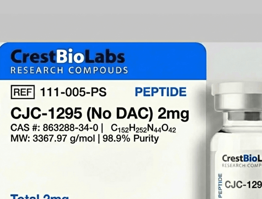
CJC-1295 (also designated as modified GRF(1-29) or tetrasubstituted GHRH(1-29)) incorporates four amino acid substitutions: D-Ala2 (DPP-IV resistance), Gln8 (metabolic stabilization), Ala15 (helix stabilization), and Leu27 (enhanced hydrophobic receptor contact). These modifications collectively extend the in-vitro half-life in plasma from approximately 10 minutes for native GHRH to over 30 minutes for CJC-1295, while maintaining full agonist activity at GHRH-R as measured by cAMP accumulation assays in CHO-GHRH-R cells.
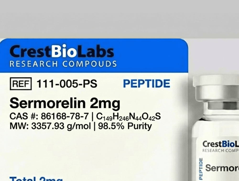
Sermorelin (GHRH(1-29)-NH2) represents the reference GHRH-R agonist for comparative SAR studies. Its C-terminal amidation mimics the naturally processed GHRH terminus and prevents carboxypeptidase degradation. Comparative receptor binding data demonstrate that sermorelin and CJC-1295 exhibit similar Ki values (low nanomolar range) at the GHRH-R, with the primary pharmacological distinction being metabolic stability rather than intrinsic receptor affinity. Truncation studies confirm that residues 30-44 of native GHRH contribute minimally to receptor binding, as GHRH(1-29) analogs retain greater than 95% of the binding affinity and functional potency of the full-length peptide in in-vitro assays (Campbell RM, et al. Peptides. 1991;12(3):569-574. doi:10.1016/0196-9781(91)90103-V).
Ghrelin Mimetics and Selective GHS-R1a Agonism
Growth hormone-releasing peptides (GHRPs) were originally developed through empirical medicinal chemistry approaches before the identification of their molecular target (GHS-R1a) and its endogenous ligand (ghrelin). The first-generation GHRPs, including GHRP-6 (His-D-Trp-Ala-Trp-D-Phe-Lys-NH2) and GHRP-2 (D-Ala-D-beta-Nal-Ala-Trp-D-Phe-Lys-NH2), were identified through structure-activity optimization of the enkephalin-derived peptide Met-enkephalin, which was found to stimulate GH release in pituitary cell cultures through a mechanism distinct from GHRH-R activation (Bowers CY. J Clin Endocrinol Metab. 2001;86(4):1464-1469. doi:10.1210/jcem.86.4.7405).
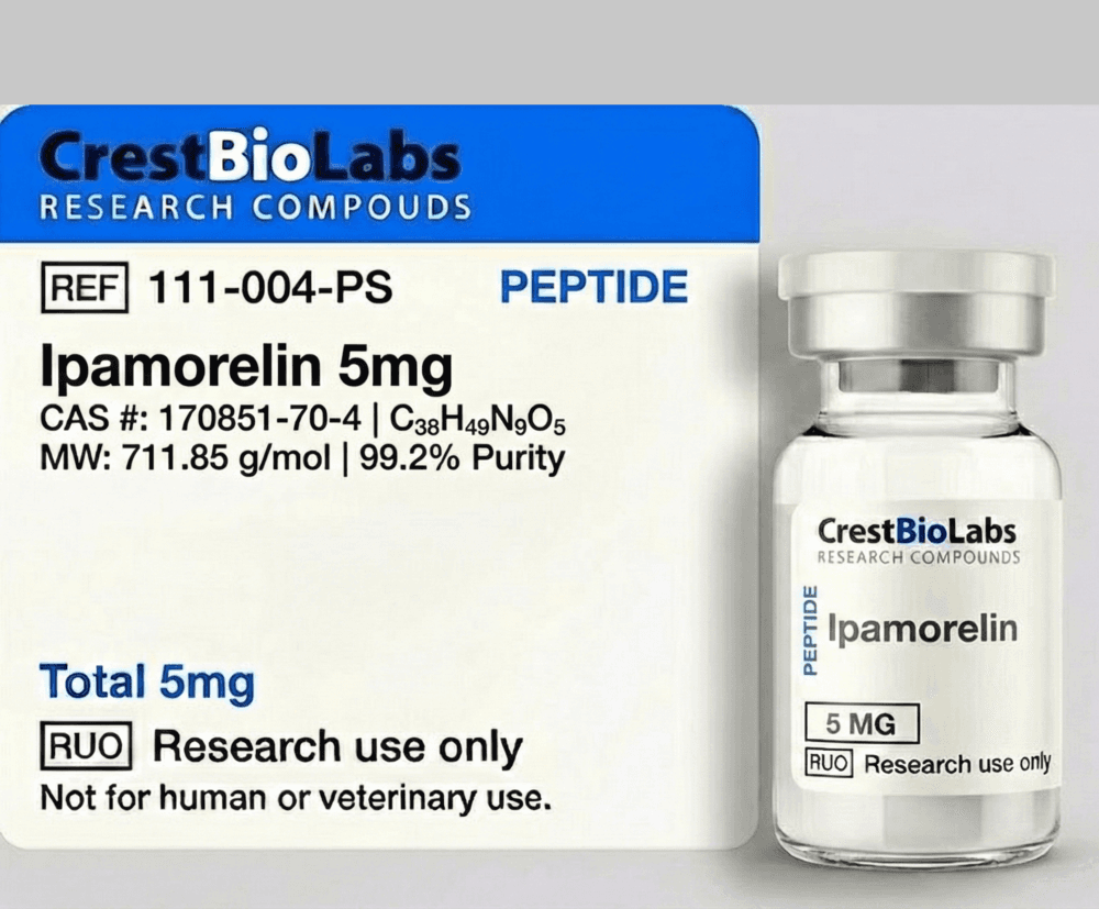
Ipamorelin emerged from a structure-activity optimization program aimed at improving GH selectivity while minimizing effects on other pituitary hormones. Its pentapeptide structure (Aib-His-D-2-Nal-D-Phe-Lys-NH2) incorporates several key design features: the Aib (alpha-aminoisobutyric acid) N-cap stabilizes helical conformation, D-2-Nal (D-2-naphthylalanine) at position 3 and D-Phe at position 4 provide optimal hydrophobic receptor contacts, and the C-terminal Lys-NH2 contributes electrostatic interactions with the receptor binding pocket. In in-vitro studies using rat pituitary cell cultures, ipamorelin was demonstrated to release GH with potency comparable to GHRP-6 while producing no statistically significant stimulation of ACTH, cortisol, or prolactin secretion at concentrations up to 100-fold above its GH-releasing EC50 (Raun K, et al. Eur J Endocrinol.1998;139(5):552-561. doi:10.1530/eje.0.1390552).
The molecular basis for ipamorelin's selectivity has been investigated using site-directed mutagenesis of GHS-R1a and computational docking studies. Crystal and cryo-EM structures of GHS-R1a in complex with various agonists have revealed that ipamorelin occupies the orthosteric binding pocket with a distinct orientation compared to less selective GHRPs: its D-2-Nal moiety engages a subpocket (defined by TM3, TM5, and ECL2 residues) that is not accessed by GHRP-6, potentially explaining the differential downstream signaling profiles. This binding mode has been hypothesized to stabilize a receptor conformation that preferentially activates Gq-mediated calcium signaling over other pathways such as ERK activation or beta-arrestin recruitment.
Comparative in-vitro profiling of GHRP-6, GHRP-2, ipamorelin, and hexarelin in GHS-R1a-expressing cell lines has established a hierarchy of selectivity for GH release over other pituitary hormones. In perifused rat pituitary cell columns, ipamorelin shows the narrowest selectivity window, stimulating GH release without affecting ACTH or prolactin secretion at concentrations up to 1 micromolar. GHRP-6 and GHRP-2 stimulate both GH and ACTH release at higher concentrations, while hexarelin activates GH, ACTH, and prolactin release in preclinical pituitary models. These selectivity profiles are critical parameters for selecting appropriate GHS-R1a agonists in in-vitro research protocols where specificity of the GH axis is required.
Synergistic Receptor Co-Activation in Pituitary Somatotroph Models
One of the most pharmacologically significant findings in GH secretagogue research is the synergistic interaction between GHRH-R and GHS-R1a signaling in pituitary somatotroph cells. When GHRH and a GHS-R1a agonist are co-administered in in-vitro or ex-vivo pituitary preparations, the resulting GH release is markedly greater than the arithmetic sum of the individual responses. This synergy was first systematically characterized using perifused rat pituitary cell preparations and has since been confirmed in primary human pituitary cell cultures (Bowers CY, et al. Endocrinology.1984;114(5):1537-1545. doi:10.1210/endo-114-5-1537).
The mechanistic basis for this synergy lies in the convergence of two distinct intracellular signaling cascades at the level of the secretory apparatus. GHRH-R activation elevates cAMP and activates PKA, which phosphorylates components of the vesicle release machinery including SNAP-25, synaptotagmin, and Munc18. Simultaneously, GHS-R1a activation via the Gq-PLC-IP3 pathway mobilizes intracellular calcium and activates PKC, which phosphorylates a complementary set of exocytotic proteins. The convergence of PKA- and PKC-mediated phosphorylation events at the SNARE complex (syntaxin-1, SNAP-25, VAMP/synaptobrevin) is hypothesized to lower the energy barrier for vesicle fusion beyond what either kinase achieves independently, producing the observed super-additive GH release.
Electrophysiological studies using perforated patch-clamp recordings from identified somatotroph cells have provided additional insight into the synergy mechanism. GHRH-mediated cAMP elevation inhibits BK (big-conductance calcium-activated potassium) channels and activates L-type calcium channels, depolarizing the membrane and increasing action potential frequency. GHS-R1a-mediated IP3-dependent calcium release from ER stores further modulates membrane potential through calcium-activated chloride channels. The combined effect produces sustained membrane depolarization and calcium oscillations with greater amplitude and frequency than either stimulus alone, as recorded in somatotroph cells from transgenic GH-GFP reporter mice.
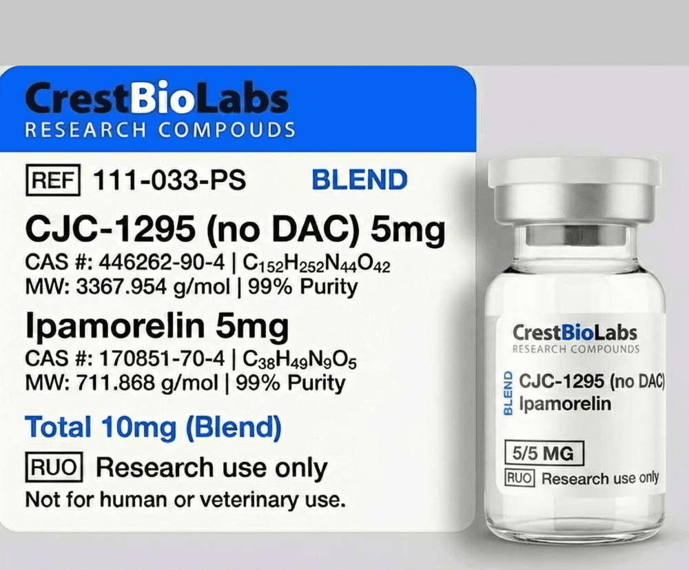
The CJC-1295 / Ipamorelin research blend is designed to enable studies of this synergistic interaction in vitro. By providing a stabilized GHRH-R agonist (CJC-1295, with DPP-IV-resistant tetrasubstituted structure) and a selective GHS-R1a agonist (ipamorelin, with established selectivity for GH release) in a single formulation, researchers can investigate the concentration-response relationships of dual-receptor co-activation using consistent molar ratios. In perifused pituitary cell assays, the synergy coefficient (measured as the ratio of combined response to the sum of individual responses) typically ranges from 2.5 to 5.0 in published studies, depending on the concentrations employed and the specific cell model used (Frohman LA, Jansson JO. Endocr Rev. 1986;7(3):223-253. doi:10.1210/edrv-7-3-223).
Receptor Binding Assays and Functional In-Vitro Models
Characterization of GHRH-R and GHS-R1a agonists requires a panel of complementary in-vitro assays that assess ligand binding affinity, functional potency, receptor selectivity, and downstream signaling profiles. The choice of assay platform and cell model significantly influences the pharmacological parameters obtained, and understanding these methodological considerations is essential for accurate interpretation of published SAR data.
GHRH-R Radioligand Binding and cAMP Assays
GHRH-R binding is typically assessed using [125I]-Tyr10-GHRH(1-44) or [125I]-His1-GHRH(1-29)-NH2 as radioligands in competition binding assays with rat anterior pituitary membranes or CHO/HEK293 cell membranes stably expressing human or rat GHRH-R. Saturation binding experiments yield equilibrium dissociation constants (Kd) and receptor density (Bmax), while competition curves provide IC50 values from which inhibition constants (Ki) are calculated using the Cheng-Prusoff equation. Functional potency at GHRH-R is measured by cAMP accumulation in GHRH-R-expressing cell lines using homogeneous time-resolved fluorescence (HTRF) or AlphaScreen-based cAMP detection kits, generating concentration-response curves with EC50 and Emax values.
GHS-R1a Binding, Calcium, and IP3 Assays
GHS-R1a binding is assessed using [125I]-ghrelin or [125I]-His-D-Trp-Ala-Trp-D-Phe-Lys-NH2 ([125I]-GHRP-6) as radioligands. Because GHS-R1a signals primarily through Gq, the preferred functional readout is intracellular calcium mobilization, measured using calcium-sensitive fluorescent indicators (Fluo-4, Fura-2) on FLIPR or plate-reader platforms. IP3 turnover assays using [3H]-myo-inositol-labeled cells provide a direct measure of PLC activation. Additionally, [35S]-GTPgammaS binding assays measure agonist-stimulated G-protein activation in cell membranes, providing a G-protein-proximal functional readout that is independent of downstream signaling amplification.
Primary Pituitary Culture and Perifusion Systems
For studying GH release under physiologically relevant conditions, primary dispersed anterior pituitary cells (typically from rat or mouse) are cultured in monolayers or perifusion columns. Perifusion systems, where cells are packed in microcolumns and continuously superfused with buffer, allow real-time measurement of pulsatile GH release in response to sequential or combined agonist stimulation. Effluent fractions are collected at 1-5 minute intervals and GH concentration is measured by radioimmunoassay (RIA) or ELISA. This system is particularly valuable for studying the temporal dynamics of synergistic GHRH + GHRP stimulation and for characterizing the kinetics of GH release and desensitization in response to sustained or repeated agonist exposure.
Selectivity Profiling and Counter-Screening
Because GHRH-R and GHS-R1a are pharmacologically distinct receptors (class B vs class A GPCRs, Gs vs Gq coupling), selectivity profiling is essential for characterizing novel agonists. Counter-screening GHRH analogs at GHS-R1a (calcium assay) and GHS-R1a agonists at GHRH-R (cAMP assay) confirms target selectivity. Additional counter-screens against related class B GPCRs (PACAP-R/PAC1, VPAC1, VPAC2, CRF-R1, glucagon receptor) and class A GPCRs (motilin receptor, neurotensin receptor) are conducted to establish broader selectivity profiles. Published selectivity data for sermorelin, CJC-1295, and ipamorelin demonstrate high target selectivity with greater than 1000-fold selectivity over the counter-screened receptors in standard in-vitro panels.