Introduction: The Importance of Standardized Handling Protocols
Research peptides are high-value, low-quantity materials that demand careful handling at every stage — from the moment a shipment arrives at the receiving dock through final use in an experiment. Unlike bulk laboratory reagents measured in grams or liters, most synthetic peptides are supplied in milligram quantities (typically 1 to 20 mg per vial), making losses from improper handling, adsorption, or contamination proportionally significant. A single mishandled reconstitution can render an entire vial unusable.
Standardized laboratory handling protocols serve multiple purposes: they maximize peptide recovery and integrity, ensure consistency between experiments and between researchers, satisfy institutional and regulatory documentation requirements, and minimize safety risks associated with handling biologically active research compounds. This article presents a comprehensive set of standard operating procedures covering the complete peptide handling workflow from shipment receipt to waste disposal.
Receiving and Inspecting Peptide Shipments
Upon receiving a peptide shipment, inspect the outer packaging for signs of damage, excessive moisture, or temperature excursion indicators (if present). Most lyophilized peptides are shipped on dry ice or with cold packs to maintain temperatures below ambient during transit. Verify that residual dry ice remains in the shipping container (for overnight shipments) or that cold packs are still cool to the touch. Document the condition of the outer package and any temperature indicators in your receiving log.
Remove the inner product vials and inspect each one individually. Check that the vial seal (crimp cap) is intact and that the stopper has not been displaced. Examine the lyophilized cake through the glass: it should appear as a white to off-white porous plug at the bottom of the vial, consistent with the product description. Document any anomalies including cake collapse, discoloration, liquid residue (meltback), or a loose or absent crimp seal. Photograph the vial contents if your receiving protocol requires visual documentation.
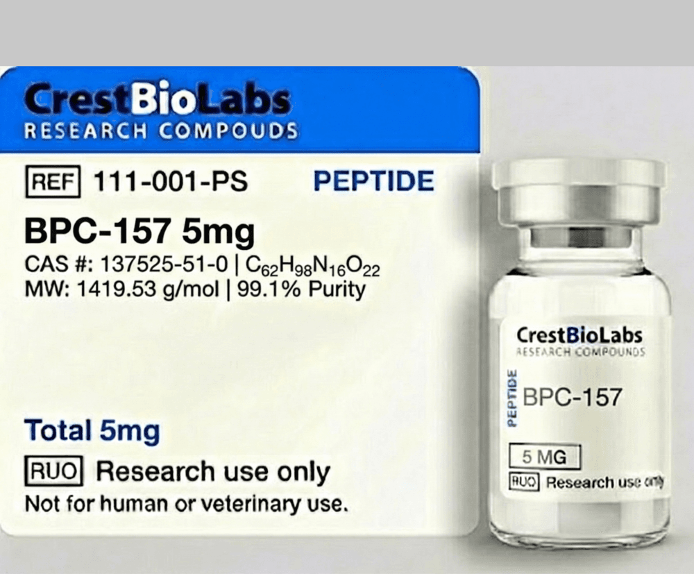
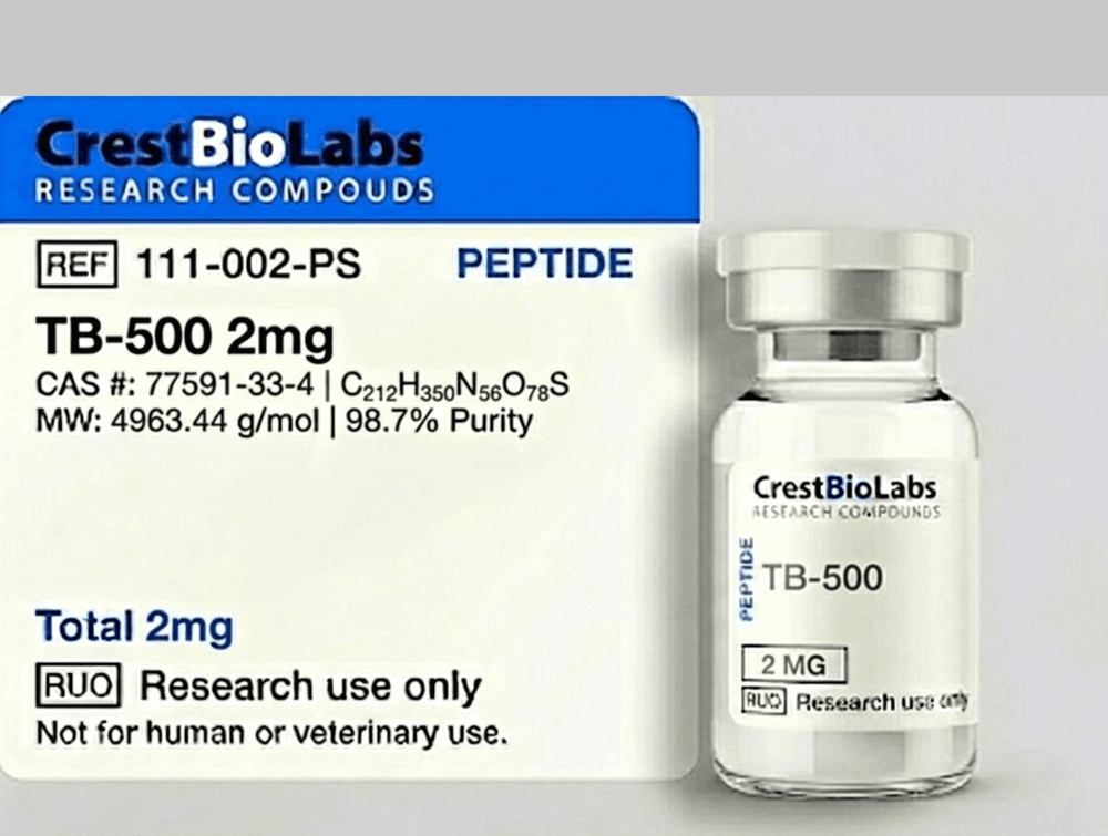
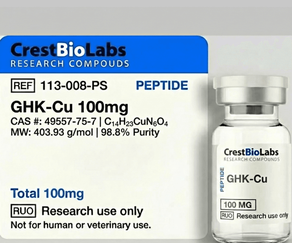
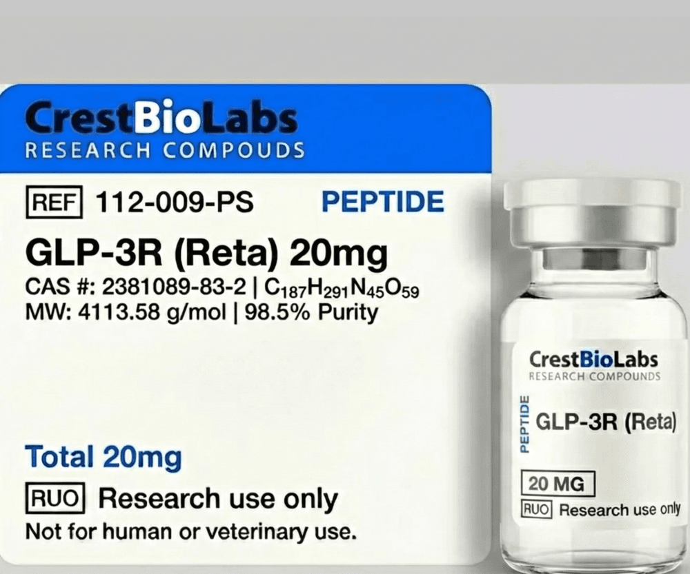
Verify the product label information against the purchase order: compound name, catalog number, lot number, quantity, and any handling or storage instructions. Locate and file the certificate of analysis (COA), which should accompany the shipment or be available for download. The COA typically includes HPLC purity data, mass spectrometry confirmation of molecular identity, and appearance description. Compare the stated purity and mass values against your procurement specifications. Once inspection is complete, transfer vials to their designated storage location (typically -20 degrees Celsius or -80 degrees Celsius) without delay.
Proper Thawing and Equilibration Procedures
When a lyophilized peptide vial is removed from frozen storage for reconstitution, it must be equilibrated to ambient room temperature before the seal is broken. This step is critical because opening a cold vial in a warm, humid laboratory environment causes immediate moisture condensation on the inner walls and the surface of the lyophilized cake. This condensed water locally dissolves the outer layer of the cake, potentially denaturing the peptide and creating a wet surface film that is difficult to incorporate uniformly during subsequent reconstitution.
Place the sealed vial on the bench and allow it to warm for 15 to 30 minutes, depending on starting temperature and vial size. Do not attempt to accelerate this process with heat sources (water baths, heat blocks, or microwaves), which create temperature gradients and may overheat the peptide. A simple equilibration at ambient temperature (20 to 25 degrees Celsius) is sufficient. During this waiting period, prepare your reconstitution solvent, calculate the required volume, and set up a clean workspace. The equilibration time can be productively used for labeling aliquot tubes and preparing your laboratory notebook entry.
Reconstitution: Solvent Selection and Technique
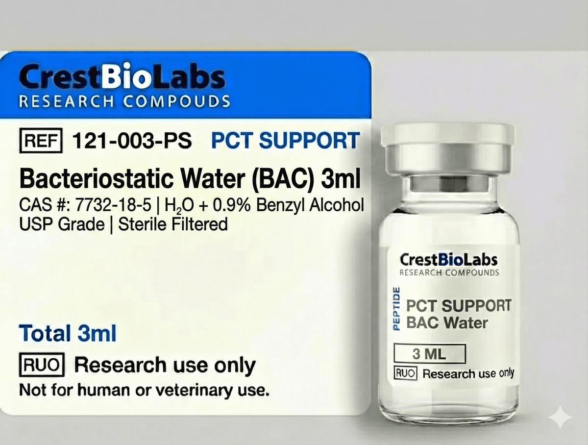
The choice of reconstitution solvent depends on the physicochemical properties of the peptide, the downstream application, and whether multi-use storage is intended. The two most common aqueous solvents are bacteriostatic water (containing 0.9% benzyl alcohol as a preservative) and sterile water for injection (preservative-free). Bacteriostatic water is preferred for multi-use vials that will be accessed over days or weeks, while sterile water is appropriate for single-use preparations or when benzyl alcohol might interfere with the assay system.
For hydrophobic peptides that resist dissolution in pure water, a two-step reconstitution protocol is often effective. First, add a small volume (50 to 100 microliters) of a solubilizing co-solvent — typically DMSO, 0.1% acetic acid, 0.1% TFA in water, or 10% acetonitrile in water — directly to the lyophilized cake. Allow this to wet and dissolve the peptide fully (1 to 2 minutes). Then dilute to the final volume with the primary aqueous solvent. This stepwise approach ensures complete dissolution of the hydrophobic peptide core before dilution into the larger aqueous volume, preventing precipitation.
When adding solvent to the vial, use a syringe fitted with an appropriate-gauge needle (typically 18 to 21 gauge). Insert the needle through the septum and direct the solvent stream gently down the inner wall of the vial, not directly onto the lyophilized cake. This minimizes foaming (which can trap peptide at the air-liquid interface and cause surface denaturation) and promotes gradual, uniform wetting of the porous cake structure.
After solvent addition, withdraw the needle and allow the vial to sit undisturbed for 3 to 5 minutes. The porous cake will gradually absorb solvent by capillary action and dissolve. Then gently swirl or roll the vial between your palms to ensure homogeneity. Do not vortex vigorously, as the mechanical shear and repeated passage of peptide through the air-liquid interface cause aggregation. If gentle swirling does not achieve complete dissolution, brief sonication in a water bath sonicator (30 to 60 seconds at room temperature) may be employed. Inspect the solution visually: it should be clear and free of visible particles.
Calculating Reconstitution Volumes and Final Concentrations
Accurate concentration determination requires accounting for the difference between gross powder weight and net peptide content. The gross weight stated on the vial label includes the peptide itself, counterions (commonly acetate or trifluoroacetate salts from HPLC purification), and residual moisture and volatiles from the lyophilization process. Net peptide content is typically 70 to 85% of the gross weight for acetate-salt peptides and 50 to 75% for TFA-salt peptides, though the exact value depends on the specific sequence and its counterion content.
For gravimetric concentration calculations: Concentration (mg/mL) = Net Peptide Mass (mg) / Reconstitution Volume (mL). If the COA provides a net peptide content percentage, multiply the label mass by this percentage to obtain the true peptide mass. For example, a 5 mg vial with 80% peptide content contains 4.0 mg of actual peptide; adding 4.0 mL of solvent yields a 1.0 mg/mL solution.
For molar concentrations: Concentration (micromolar) = [Net Peptide Mass (mg) x 1000] / [Molecular Weight (g/mol) x Volume (mL)]. Using the same example with a peptide of molecular weight 1420 g/mol: (4.0 x 1000) / (1420 x 4.0) = 0.704 millimolar, or 704 micromolar. When preparing serial dilutions from the stock solution, always use calibrated volumetric pipettes and make dilutions in the same buffer system to avoid pH shifts or ionic strength changes that might affect peptide solubility or activity.
Aseptic Technique and Contamination Prevention
Microbial contamination of reconstituted peptide solutions is a persistent risk in laboratory settings, particularly for solutions that will be stored and accessed over multiple days. Bacteria and fungi can metabolize the peptide as a carbon and nitrogen source, producing degradation products that interfere with downstream assays and introducing endotoxins that can confound cell-based experiments. Even a single contamination event can render the entire reconstituted supply unusable.
To minimize contamination risk, perform all reconstitution and aliquoting operations in a clean environment — ideally a laminar flow hood or biosafety cabinet. Wipe the work surface with 70% ethanol or isopropanol before and after use. Swab the vial septum with an alcohol wipe before each needle insertion. Use individually wrapped, sterile, disposable syringes and needles; never reuse a syringe. For pipetting operations, use sterile, filtered pipette tips.
If working outside a laminar flow environment, minimize the time that vials and tubes are open. Have all tubes pre-labeled and arranged in a rack before beginning the aliquoting process. Work quickly and close tubes immediately after dispensing. The use of bacteriostatic water as the reconstitution solvent provides a degree of antimicrobial protection, but it does not substitute for proper aseptic technique — benzyl alcohol at 0.9% inhibits growth but does not sterilize a contaminated solution.
Working with Milligram-Scale Quantities: Pipetting and Weighing
Research peptides are typically supplied in quantities of 1 to 20 milligrams, and individual experiments may require only micrograms of material. At these scales, losses from surface adsorption, pipetting imprecision, and weighing error become proportionally significant. Adopting best practices for small-volume and small-mass handling is essential for maximizing the utility of each vial.
For pipetting volumes below 10 microliters, use a calibrated P2 or P10 micropipette with low-retention filter tips. Pre-wet the tip by aspirating and dispensing the solution 2 to 3 times before taking the final measurement; this coats the inner tip surface and reduces volume error due to adsorption. For volumes below 1 microliter, positive-displacement pipettes are preferred over air-displacement pipettes, as they eliminate the dead volume and surface tension effects that cause inaccuracy at sub-microliter scales.
If weighing lyophilized peptide is required (for example, to subdivide a bulk quantity), use an analytical balance with a readability of 0.01 mg (10 micrograms) or better. Tare the balance with the receiving container in place, allow the reading to stabilize for at least 30 seconds, and record the weight. Perform weighing in a draft-free environment — even small air currents can cause milligram-level fluctuations on an analytical balance. For hygroscopic peptides, perform weighing quickly and in a low-humidity environment to minimize moisture uptake during the weighing process.
Documentation, Record Keeping, and Traceability
Comprehensive documentation of peptide handling is not merely administrative overhead — it is a fundamental component of reproducible science. When an unexpected result occurs in an experiment, the ability to trace the history of every reagent used — including the peptide — is essential for troubleshooting. Documentation also satisfies the requirements of institutional review boards, funding agencies, and regulatory authorities for transparent accounting of research materials.
A complete peptide handling record should include the following minimum elements: certificate of analysis (archived with lot number and purity data); purchase order and receiving date; condition upon receipt (visual inspection notes); storage location and temperature; reconstitution date, solvent, volume, and calculated concentration; aliquot map (number of aliquots, volume per aliquot, storage location); freeze-thaw history for each aliquot (date and reason for each thaw); experimental use log (date, experiment ID, volume withdrawn, remaining volume); and disposal date and method. Electronic laboratory notebook systems facilitate this tracking and provide backup, search, and audit trail capabilities that paper notebooks cannot match.
Waste Disposal and Safety Considerations
Research peptide waste — including expired or degraded peptide solutions, used vials, contaminated pipette tips, and PPE — must be disposed of in accordance with institutional environmental health and safety (EHS) policies and applicable local, state, and federal regulations. In most institutional settings, aqueous peptide solutions at research concentrations (microgram to milligram per milliliter) may be disposed of through the chemical waste stream or, for sufficiently dilute solutions, through the sanitary sewer with appropriate dilution, depending on institutional policy.
Sharps (needles, broken glass vials) must be placed in approved sharps containers. Empty glass vials that contained lyophilized peptide may be disposed of as regular glass waste after confirming that no residual powder remains, or as chemical waste if the institutional policy requires it. Organic solvents (DMSO, acetonitrile, acetic acid) used in reconstitution must be collected in appropriate waste containers and disposed of through the hazardous waste program.
Regarding personal safety, all research peptides should be handled with the assumption that they are biologically active compounds of incompletely characterized hazard. Always wear nitrile gloves, a laboratory coat, and eye protection when handling peptides. Avoid generating aerosols: do not aggressively vortex open tubes, and exercise caution when handling lyophilized powder, which can become airborne due to static charge and low particle density. If a powder spill occurs, wet the affected area with water or damp wipes before cleaning — do not attempt to sweep dry powder, as this generates a dust plume. Always consult the Safety Data Sheet (SDS) for each specific compound for substance-specific hazard information, first-aid measures, and spill response procedures.