Classification of Cosmetic Peptides in Dermatological Research
Peptides investigated in dermatological research are classified according to their primary mechanism of action in in-vitro and preclinical models. This functional classification system provides a framework for understanding how different peptide sequences interact with skin biology at the molecular level, and guides the selection of appropriate assays for characterizing their activity.
Signal peptides are sequences that modulate cellular gene expression, typically by interacting with cell surface receptors or intracellular signaling molecules. In fibroblast culture models, signal peptides have been investigated for their ability to stimulate the production of extracellular matrix (ECM) proteins including type I collagen, type III collagen, elastin, and fibronectin. The prototypical signal peptide studied in research contexts is the pentapeptide KTTKS (Lys-Thr-Thr-Lys-Ser), a fragment of the type I procollagen C-proteinase recognition sequence.
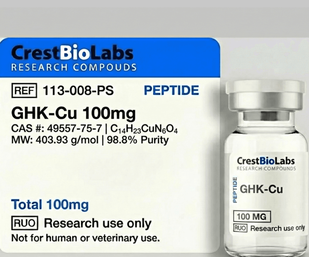
Carrier peptides function as delivery vehicles for biologically active metal ions, particularly copper(II). The copper-binding tripeptide GHK-Cu is the most extensively studied carrier peptide, facilitating the intracellular delivery of copper to support copper-dependent enzymatic processes including lysyl oxidase activity, superoxide dismutase function, and cytochrome c oxidase-mediated mitochondrial respiration.
Enzyme-inhibitor peptides are sequences designed to modulate protease activity in skin tissue models. Matrix metalloproteinases (MMPs) and other proteases play central roles in ECM turnover, and peptide-based inhibitors of these enzymes have been investigated in preclinical models for their effects on collagen degradation rates. Neurotransmitter-affecting peptides, the fourth category, have been studied in in-vitro neuromuscular junction models for their effects on acetylcholine release and SNARE complex assembly.
Copper Peptide Biology: The GHK-Cu Mechanism
GHK-Cu (glycyl-L-histidyl-L-lysine copper(II) complex) is a naturally occurring tripeptide-metal complex first identified in human plasma by Dr. Loren Pickart in 1973. The peptide is present in plasma at concentrations of approximately 200 ng/mL in young adults, declining to approximately 80 ng/mL by age 60. This age-related decline in circulating GHK-Cu concentration has been a subject of interest in preclinical aging research.
The copper(II) binding site of GHK involves a square-planar coordination geometry. The metal ion is chelated by the alpha-amino group of glycine, the amide nitrogen of the Gly-His peptide bond (after deprotonation), the N-pi nitrogen of the histidine imidazole ring, and typically a water molecule or carboxylate oxygen in the fourth coordination position. This coordination complex has a remarkably high binding affinity (log K = 16.44 at pH 7.4), ensuring that GHK effectively sequesters and transports copper(II) under physiological conditions investigated in in-vitro studies.
Gene expression profiling studies using microarray analysis have demonstrated that GHK-Cu modulates the expression of over 4,000 human genes at concentrations of 1-10 micromolar in cultured fibroblasts. The upregulated gene networks include those involved in collagen synthesis (COL1A1, COL3A1), glycosaminoglycan production, nerve growth factor signaling, and antioxidant defense systems (superoxide dismutase, glutathione-related genes). Simultaneously, GHK-Cu has been observed to downregulate pro-inflammatory cytokine expression (IL-6, TNF-alpha) and fibrinogen synthesis in in-vitro models.
In cell culture wound healing assays (scratch assays), GHK-Cu has been investigated for its effects on fibroblast migration, proliferation, and collagen deposition. Published in-vitro data suggest that GHK-Cu stimulates the secretion of vascular endothelial growth factor (VEGF) and basic fibroblast growth factor (bFGF), which are key mediators of angiogenesis in tissue remodeling models. The peptide also modulates metalloproteinase activity, increasing MMP-2 (gelatinase A) while suppressing certain inflammatory MMPs, suggesting a role in coordinated matrix remodeling rather than simple degradation or synthesis.
Melanotropic Peptides and MC1R Signaling
Melanotropic peptides are analogs of alpha-melanocyte-stimulating hormone (alpha-MSH), a 13-amino acid peptide derived from post-translational processing of proopiomelanocortin (POMC) in the pituitary gland and skin. Alpha-MSH signals primarily through the melanocortin-1 receptor (MC1R) expressed on epidermal melanocytes, activating the cAMP/PKA signaling cascade that drives melanogenesis, the biosynthesis of melanin pigments.
The melanogenesis pathway proceeds through a well-characterized enzymatic cascade in melanocytes. MC1R activation elevates intracellular cAMP, which activates CREB (cAMP response element-binding protein) and subsequently upregulates MITF (microphthalmia-associated transcription factor) expression. MITF transcriptionally activates the genes encoding tyrosinase (TYR), tyrosinase-related protein-1 (TYRP1), and dopachrome tautomerase (DCT/TYRP2), the three key enzymes in the melanin synthesis pathway. Tyrosinase catalyzes the rate-limiting step: the hydroxylation of L-tyrosine to L-DOPA and subsequent oxidation to dopaquinone, which then undergoes either cyclization to form eumelanin (brown-black pigment) or conjugation with cysteine to form pheomelanin (red-yellow pigment).
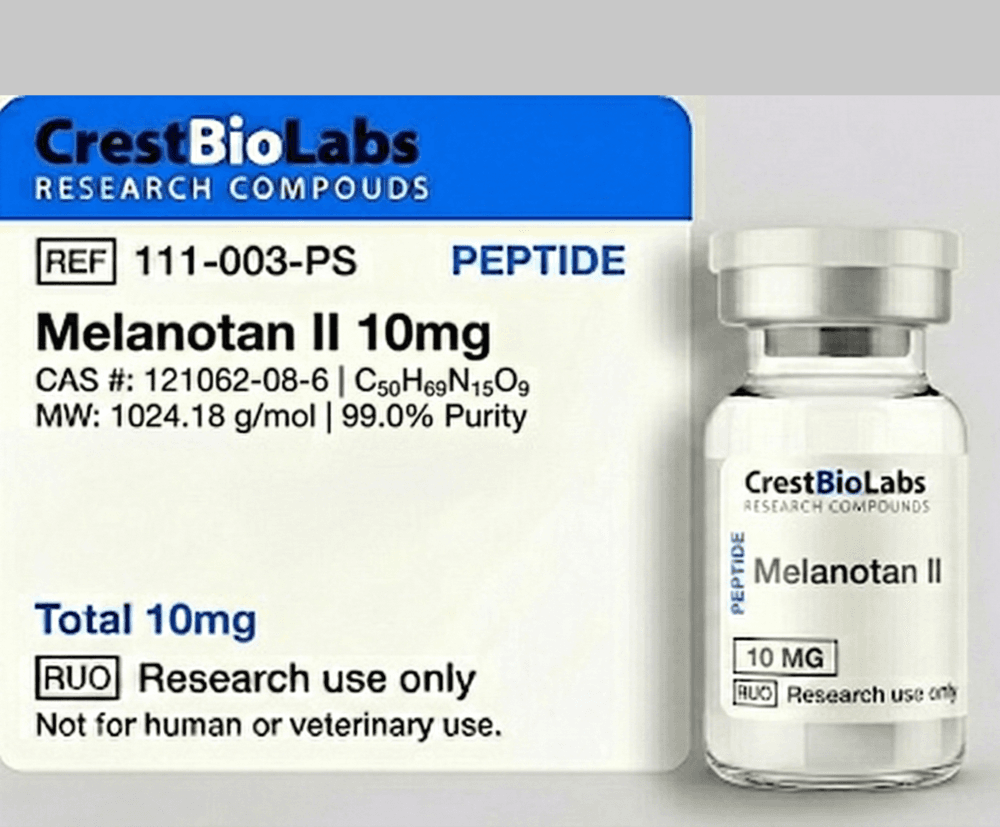
Synthetic alpha-MSH analogs such as Melanotan II (Ac-Nle-cyclo[Asp-His-D-Phe-Arg-Trp-Lys]-NH2) are cyclic peptides that exhibit enhanced potency and stability compared to native alpha-MSH. The D-phenylalanine substitution at position 7 and cyclization through a lactam bridge confer resistance to enzymatic degradation and improved receptor binding affinity. Melanotan II functions as a non-selective agonist at melanocortin receptors MC1R through MC5R, and its melanogenic activity has been characterized in B16 murine melanoma cells, primary human melanocyte cultures, and three-dimensional skin equivalent models.
In-vitro assays for characterizing melanotropic peptide activity include melanin content quantification (spectrophotometric measurement of NaOH-solubilized melanin at 475 nm), tyrosinase activity assays (L-DOPA oxidation kinetics), MC1R binding assays using radiolabeled [Nle4,D-Phe7]-alpha-MSH, and fluorescence-based cAMP accumulation assays in MC1R-expressing cell lines. These in-vitro methods provide a comprehensive characterization of melanotropic peptide pharmacology under controlled laboratory conditions.
Collagen-Stimulating Peptides in Research
Collagen constitutes approximately 70-80% of the dry weight of the dermis, with type I collagen comprising roughly 80% and type III collagen approximately 15% of total dermal collagen. The rate of collagen synthesis by dermal fibroblasts decreases with age in preclinical models, while collagen degradation by matrix metalloproteinases (particularly MMP-1, MMP-3, and MMP-9) increases, resulting in a net collagen deficit that has been quantified in ex-vivo skin biopsies and in-vitro fibroblast aging models.
The pentapeptide KTTKS is derived from the C-terminal propeptide of type I procollagen, specifically from the sequence recognized by the C-proteinase BMP-1 during procollagen processing. In-vitro studies using human dermal fibroblast cultures have investigated the ability of KTTKS and its palmitoylated derivative (palmitoyl pentapeptide-4, or Pal-KTTKS) to stimulate type I and type III procollagen synthesis, as well as fibronectin production. The palmitoyl modification enhances membrane permeability in cell culture models by increasing the peptide's lipophilicity and facilitating interaction with the lipid bilayer of the cell membrane.
Matrikines represent a broader class of ECM-derived peptides that are generated during matrix remodeling by protease activity and subsequently modulate cellular behavior. The concept of matrikine signaling provides a theoretical framework for understanding how collagen fragments and other ECM-derived peptides can influence fibroblast activity, migration, and matrix protein production through feedback loops in in-vitro tissue models.
Research into collagen-stimulating peptides employs multiple quantitative endpoints including procollagen type I C-peptide (PICP) ELISA, hydroxyproline content assays, Sirius red staining for total collagen visualization, and quantitative PCR for COL1A1 gene expression. Three-dimensional collagen lattice contraction assays provide additional functional data by measuring fibroblast-mediated contraction of reconstituted collagen gels in the presence or absence of test peptides.
Signal Peptides, Carrier Peptides, and Enzyme-Inhibitor Peptides
The functional classification of cosmetic peptides into signal, carrier, and enzyme-inhibitor categories reflects distinct molecular mechanisms through which these sequences interact with skin biology. Understanding these mechanistic differences is essential for designing appropriate in-vitro assay strategies and interpreting preclinical data.
Signal Peptides: Receptor-Mediated Gene Modulation
Signal peptides modulate cellular behavior through direct interaction with cell surface receptors or intracellular targets. TGF-beta mimetic peptides, for example, have been investigated for their ability to activate the TGF-beta/Smad signaling pathway in fibroblast cultures, leading to upregulation of collagen and elastin gene expression. The hexapeptide VGVAPG (Val-Gly-Val-Ala-Pro-Gly), a repetitive motif from elastin, has been studied for its interaction with the elastin-binding protein (EBP/S-Gal) and its downstream effects on matrix protein production in dermal fibroblast models.
Carrier Peptides: Metal Ion Transport
Beyond GHK-Cu, other metal-binding peptides have been investigated in dermatological research. Manganese-binding peptides and zinc-binding sequences have been studied for their ability to deliver these essential trace elements to cells in culture, supporting metal-dependent enzymatic processes including superoxide dismutase (SOD) activity and zinc finger transcription factor function. The specificity of metal coordination chemistry determines which metal ion each carrier peptide preferentially binds and delivers.
Enzyme-Inhibitor Peptides: Protease Modulation
Matrix metalloproteinase (MMP) inhibitory peptides are investigated for their ability to reduce collagen degradation rates in in-vitro models. These peptides typically contain sequences that mimic the MMP cleavage site on collagen substrates, functioning as competitive inhibitors. Tissue inhibitor of metalloproteinase (TIMP)-derived peptide fragments and synthetic hydroxamate-containing peptides have been evaluated in fluorogenic MMP activity assays and collagen degradation assays. The balance between MMP activity and TIMP inhibition is a key parameter in ECM homeostasis models relevant to skin research.
Skin Barrier Function and Peptide Interactions
The stratum corneum, the outermost layer of the epidermis, constitutes the primary physical barrier of the skin. It consists of terminally differentiated keratinocytes (corneocytes) embedded in a highly organized lipid matrix composed of ceramides, cholesterol, and free fatty acids arranged in lamellar bilayer structures. This "brick and mortar" architecture presents a significant barrier to the penetration of hydrophilic molecules, including most peptides.
Transepidermal water loss (TEWL) is a standard in-vitro and in-vivo parameter used to assess skin barrier integrity. Certain peptides have been investigated in preclinical models for their effects on barrier function. Antimicrobial peptides (AMPs) such as human beta-defensins and cathelicidin LL-37 play dual roles in barrier biology, functioning both as innate immune effectors and as regulators of keratinocyte differentiation and barrier repair processes in in-vitro wound models.
Research into peptide-barrier interactions encompasses both the effects of peptides on barrier function and the barrier's influence on peptide penetration. Ceramide-mimetic peptides and peptides that stimulate ceramide synthase expression have been investigated for their ability to enhance barrier function in reconstructed human epidermis models. Conversely, certain cell-penetrating peptides (CPPs) such as the TAT peptide (GRKKRRQRRRPQ) and penetratin (RQIKIWFQNRRMKWKK) have been studied for their ability to traverse the stratum corneum and facilitate the intracellular delivery of conjugated bioactive cargo.
Tight junction proteins (claudins, occludin, ZO-1) in the granular layer of the epidermis contribute to the paracellular barrier, and certain peptides have been investigated for their effects on tight junction assembly and function in keratinocyte monolayer models. Understanding these barrier interactions at the molecular level is essential for predicting the bioavailability and biological activity of topically applied peptides in research settings.
In-Vitro Skin Models for Peptide Research
The evaluation of cosmetic peptide activity relies on a hierarchy of in-vitro models that range from simple monolayer cultures to complex three-dimensional tissue equivalents. Each model system offers distinct advantages and limitations that influence experimental design and data interpretation.
Two-Dimensional Cell Culture Models
Monolayer cultures of primary human dermal fibroblasts (HDFs) are the most widely used system for evaluating collagen-stimulating and ECM-modulating peptides. HDFs can be cultured from neonatal or adult skin biopsies and maintain their ability to synthesize type I and type III collagen, elastin, fibronectin, and glycosaminoglycans for multiple passages. Primary human epidermal keratinocytes (NHEKs) are used for barrier function studies, differentiation assays, and inflammatory response models. Melanocyte cultures, including primary human epidermal melanocytes (NHEMs) and the B16-F10 murine melanoma cell line, serve as standard models for melanogenesis research.
Three-Dimensional Reconstructed Skin Models
Reconstructed human epidermis (RhE) models consist of stratified, differentiated keratinocytes grown at the air-liquid interface on polycarbonate or collagen-coated inserts. Commercially available models include EpiDerm (MatTek), SkinEthic RHE (Episkin), and LabCyte EPI-MODEL. These models exhibit a fully differentiated epidermis with functional stratum corneum and are validated for barrier function assessment, irritation testing, and permeation studies. Full-thickness skin equivalents add a dermal compartment containing fibroblasts in a collagen or fibrin matrix, enabling studies of dermal-epidermal interactions and matrix remodeling.
Ex-Vivo Skin Explant Models
Human skin explants obtained from surgical procedures (abdominoplasty, breast reduction) maintain the full complexity of native skin tissue architecture including all cell types, appendageal structures, vascular networks, and extracellular matrix organization. Explants can be maintained in culture for 7-14 days and provide the most physiologically relevant model for evaluating peptide effects on intact skin tissue. However, donor variability, limited availability, and the progressive loss of tissue viability over time are acknowledged limitations of this model system in research applications.
Bioavailability and Delivery Considerations
The primary challenge in dermatological peptide research is achieving sufficient concentration of the active peptide at the target site within the skin. The stratum corneum's lipophilic barrier preferentially permits the passage of small (molecular weight less than 500 Da), moderately lipophilic (log P of 1-3) molecules. Most bioactive peptides exceed these parameters, being both hydrophilic and larger than the 500 Da threshold, necessitating delivery strategies that enhance skin penetration in preclinical models.
Lipidation, the conjugation of fatty acid chains (typically C14-C18 palmitic or stearic acid) to peptide N-termini or lysine side chains, is a widely investigated strategy for improving peptide skin permeation. The lipid moiety increases the peptide's partition coefficient, facilitating interaction with the lipid-rich intercellular matrix of the stratum corneum. In-vitro Franz diffusion cell experiments using excised human or porcine skin have demonstrated that palmitoylation can increase peptide permeation by 2-10 fold compared to unconjugated peptides, depending on the specific sequence and formulation vehicle.
Nanoparticle-based delivery systems represent another approach investigated in preclinical dermatological research. Liposomes, solid lipid nanoparticles (SLNs), nanostructured lipid carriers (NLCs), and polymeric nanoparticles (PLGA, chitosan) have been evaluated as carriers for cosmetic peptides. These nanocarriers can enhance skin permeation by acting as penetration enhancers, forming occlusive films, fusing with stratum corneum lipids, or accumulating in hair follicle openings (the transfollicular route). Particle size, zeta potential, lipid composition, and encapsulation efficiency are critical parameters that influence delivery performance in in-vitro skin models.
Physical enhancement techniques including iontophoresis (electrically driven transport), sonophoresis (ultrasound-mediated delivery), and microneedle arrays (creating transient micropores in the stratum corneum) have been investigated as adjunctive approaches for peptide delivery in preclinical settings. Each technique offers distinct advantages for specific peptide characteristics: iontophoresis is effective for charged peptides, sonophoresis transiently disrupts lipid bilayer structure, and microneedles physically bypass the stratum corneum barrier entirely. The choice of delivery strategy in research protocols depends on the specific peptide's physicochemical properties, the target skin compartment, and the experimental objectives of the study.